
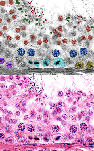
Human testicle, light micrographs. The bottom micrograph shows a seminiferous tubule. In the top micrograph the cell types of the male germinal epithelium have been marked with colour. Seen are; Sertoli cells (yellow), spermatogonia (pink), spermatogonia in metaphase (light blue), primary spermatocytes in pachytene phase (blue), spermatids (brown), spermatozoa (red) and residual bodies (green).
| px | px | dpi | = | cm | x | cm | = | MB |
Details
Creative#:
TOP27890818
Source:
達志影像
Authorization Type:
RM
Release Information:
須由TPG 完整授權
Model Release:
N/A
Property Release:
N/A
Right to Privacy:
No
Same folder images:
 Loading