
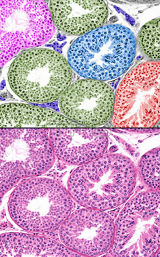
Human testicle, light micrographs. The bottom micrograph shows seminiferous tubules. The development of spermatogenesis is asynchronous among seminiferous tubules and even within zones of a single seminiferous tubule. For this reason, in histological sections of the testis, seminiferous tubules can be seen in different phases of spermatogenesis. In the top micrograph, these different phases have been marked with different colours. Amongst the seminiferous tubules are Leydig cells (purple).
| px | px | dpi | = | cm | x | cm | = | MB |
Details
Creative#:
TOP27890832
Source:
達志影像
Authorization Type:
RM
Release Information:
須由TPG 完整授權
Model Release:
N/A
Property Release:
N/A
Right to Privacy:
No
Same folder images:
 Loading