
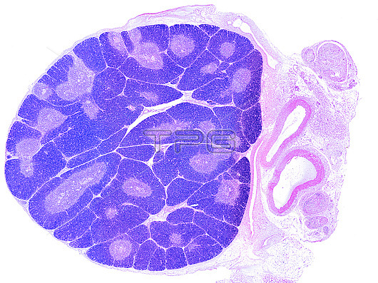
Light micrograph showing a young thymus. The organization into lobules is clearly seen. In each lobule, the peripheral cortex appears more stained, due to the high density of T-lymphocyte precursor cells. In the paler centre of each lobule there are many Hassall's corpuscles. On the right side of thymus there are blood vessels and nerves.
| px | px | dpi | = | cm | x | cm | = | MB |
Details
Creative#:
TOP28464176
Source:
達志影像
Authorization Type:
RM
Release Information:
須由TPG 完整授權
Model Release:
N/A
Property Release:
N/A
Right to Privacy:
No
Same folder images:
 Loading