
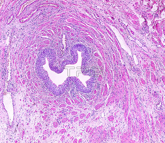
Light micrograph of a cross-sectioned human ureter stained with haematoxylin and eosin. Lining the lumen is the folded mucosa, formed by a transitional epithelium, or urothelium, with several layers of cells and the connective tissue lamina propria. Surrounding this are the muscular layers formed by fascicles of smooth muscle fibres that, unlike in the digestive tube, are not clearly arranged in layers.
| px | px | dpi | = | cm | x | cm | = | MB |
Details
Creative#:
TOP28464192
Source:
達志影像
Authorization Type:
RM
Release Information:
須由TPG 完整授權
Model Release:
N/A
Property Release:
N/A
Right to Privacy:
No
Same folder images:
 Loading