
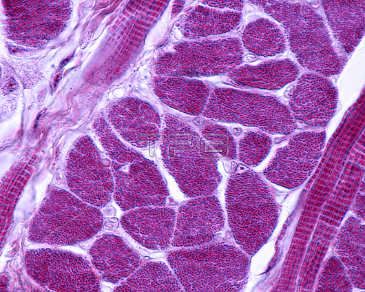
Light micrograph of a cross-sectioned skeletal muscle stained with Mallory's phosphotungstic acid haematoxylin (PTAH). Each oval or polygonal structure corresponds to a cross-sectioned myocyte. Inside each one there is a dotted pattern that occupies the entire cytoplasm that corresponds to the set of myofibrils. The nuclei, which are poorly stained with this technique, are displaced to the periphery. The narrow space between that separates some fibres from others is the endomysium, where blood capillaries are found.
| px | px | dpi | = | cm | x | cm | = | MB |
Details
Creative#:
TOP28927701
Source:
達志影像
Authorization Type:
RM
Release Information:
須由TPG 完整授權
Model Release:
n/a
Property Release:
n/a
Right to Privacy:
No
Same folder images:
 Loading