
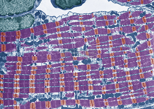
Striated muscle. Coloured transmission electron micrograph (TEM) of a longitudinal section through striated skeletal muscle. The striated banding-pattern of the muscle fibrils is seen. The fibrils run in parallel and between them runs sarcoplasmic reticulum (SR) that transmits nerve impulses to the fibrils. Here, the SR contains mitochondria (blue). Within each fibril are contractile units called sarcomeres. A sarcomere has protein filaments of myosin and actin that slide over each other, thereby causing the whole muscle to contract. Skeletal muscle is responsible for voluntary muscle movement in the body. Magnification: x10, 000 when printed at 10 centimetres wide
| px | px | dpi | = | cm | x | cm | = | MB |
Details
Creative#:
TPG32286123
Source:
達志影像
Authorization Type:
RF
Release Information:
須由TPG 完整授權
Model Release:
N/A
Property Release:
N/A
Right to Privacy:
No
Same folder images:
 Loading