
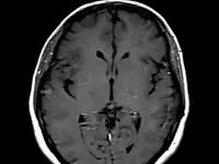
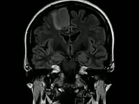
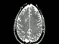
Slipped lumbar disc. Sequence of magnetic resonance imaging (MRI) sagittal scans showing the internal structure in the lower back of a 30-year-old woman with a prolapsed (slipped) intervertebral disc. In this vertical view from the side, the front of the body is at left, with the spine down right of centre. The sequence moves through the body from one side to the other, with the backbone, vertebrae (grey blocks), intervertebral discs (dark grey), cerebrospinal fluid (white), and spinal cord (grey) clearly visible in the middle of the sequence. The indentation into the spinal cord by the slipped disc is seen mid-clip at lower right at the L4/L5 level. The slipped disc is indenting the thecal sac of the cauda equina (a bundle of spinal nerves) and is compressing the nerve roots of the cauda equina. This has caused severe and sudden (acute) back pain, loss of bladder sensation, and urinary incontinence.


Details
WebID:
C00725895
Clip Type:
RM
Super High Res Size:
1920X1080
Duration:
00:00:08.000
Format:
QuickTime
Bit Rate:
24 fps
Available:
download
Comp:
200X150 (0.00 M)
Model Release:
NO
Property Release
No
 Loading